Endoscopic tympanoplasty - step by step - Prof. Marchioni and his equipe - University Hospital of Verona - Italy
Endoscopic tympanoplasty - step by step
VIDEO

PIEZOSURGERY® use
| RECOMMENDED INSERTS | |
Downloads
STEP BY STEP PROCEDURE
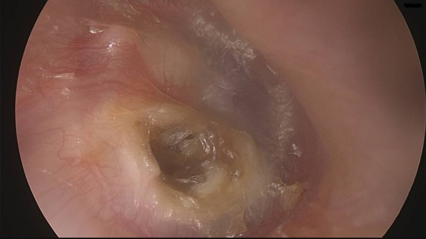
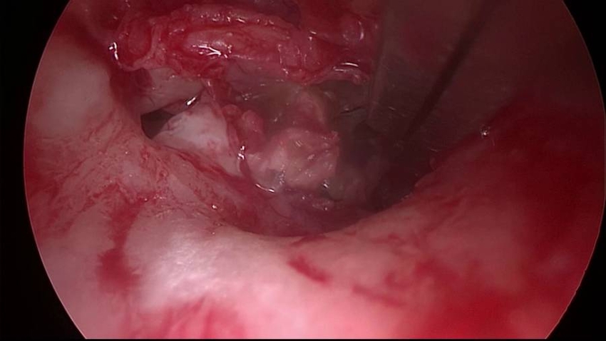
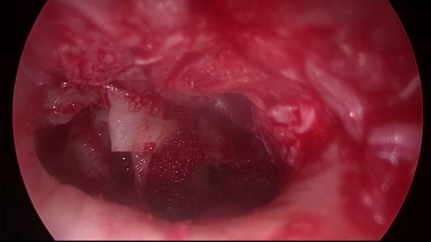
Right ear. Endoscopic view with a 0-degree endoscope. A posterior mesotympanic cholesteatoma (primary acquired) and a whitish mass medial to the tympanic membrane, anterior to the malleus are showed.
The external auditory canal is injected posteriorly.

The tympanomeatal flap is started with incision made from the 6 to 12 o’clock positions with the round knife.
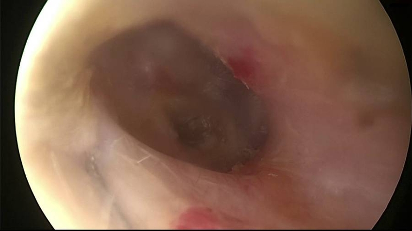
The tympanomeatal flap is raised and anteriorly pushed until reaching the annulus anteriorly. The annulus is elevated superiorly with the Rosen needle, showing the chorda tympani and the mesotympanic cholesteatoma sac.
The tympanomeatal flap is pushed forward, exposing the tympanic cavity and the cholesteatoma sac.
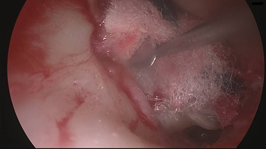
Inflammatory webs and scar tissue are removed using micro scissors.
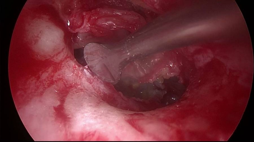
Adequate exposure of the cholesteatoma sac are achieved by removing the bone posteriorly with PIEZOSURGERY®.
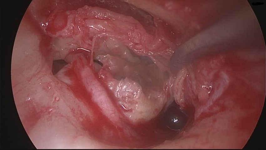
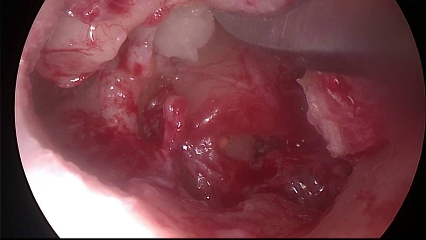
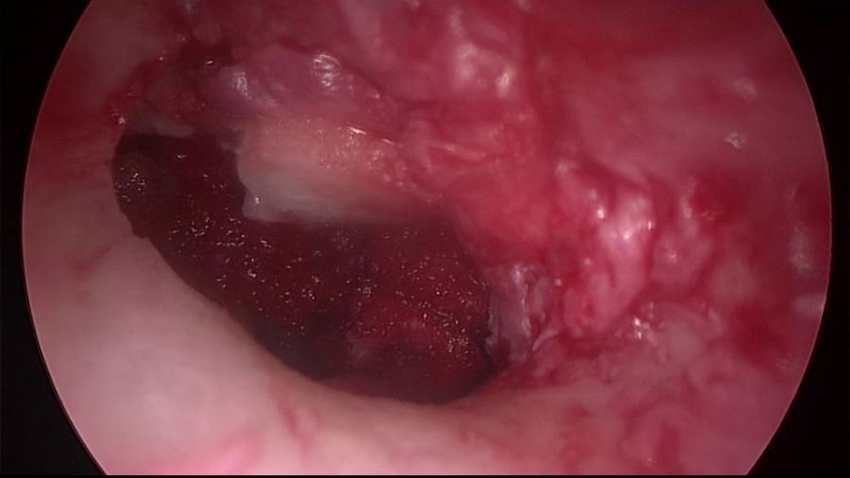
The mesotympanic and retrotympanic cholesteatoma is removed.

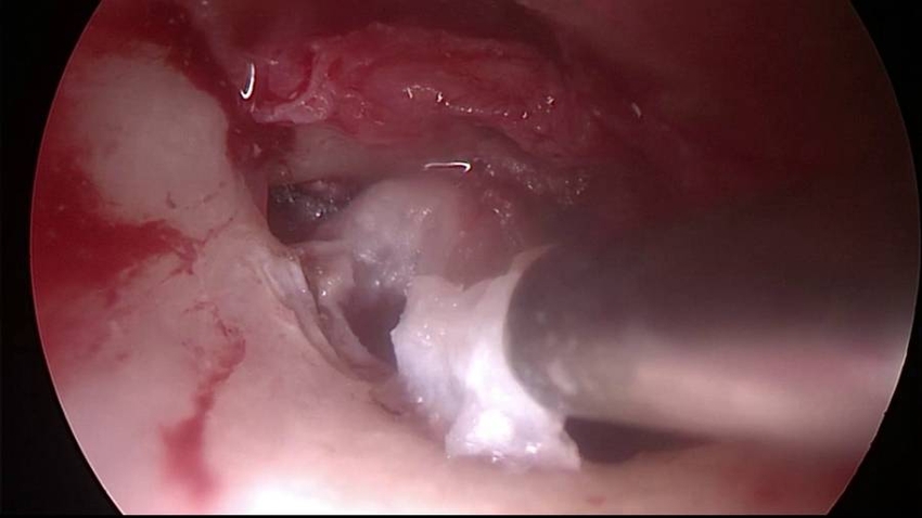
Atticotomy is performed using PIEZOSURGERY®.
The incus presents an erosion of the long process. The residual incus is removed.
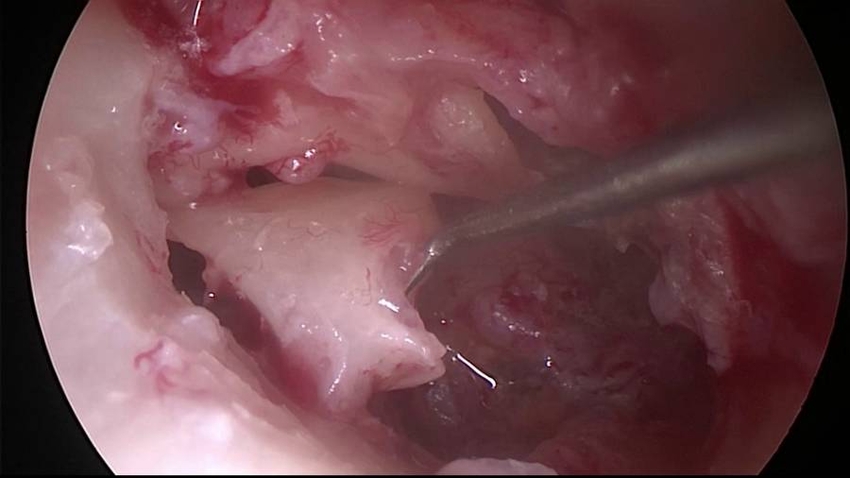
The cholesteatoma sac located anterior to the malleus is also removed.
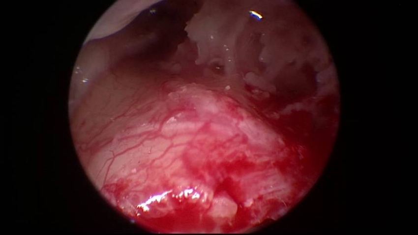
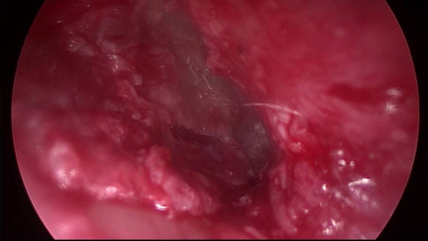
Endoscopic check of the final surgical cavity is performed using the 45-degree endoscope. The tympanic orifice of the Eustachian tube and the whole protympanic space are free from residual cholesteatoma.
Endoscopic check of the final surgical cavity with the 45-degree endoscope. The attic is free from residual cholesteatoma.
Endoscopic check of the final surgical cavity with the 45-degree endoscope. The retrotympanum is free from residual cholesteatoma.

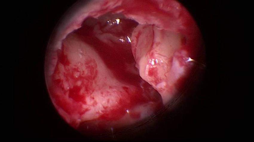
A partial ossicular replacement prosthesis (PORP) is carried out with remodeled autologous incus. A depression on the under-surface of the prosthesis is made with a small diamond bur (1 mm-diameter), in such a way that the stapes capitulum fits into it.
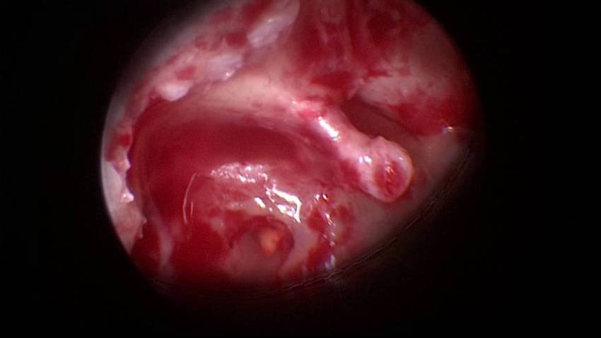
The PORP is placed over the stapes capitulum and stabilized with hemostat absorbable material (i.e. Spongostan).

The reconstruction of the tympanic membrane is performed using tragal cartilage and perichondrium graft.
Hemostat absorbable material (i.e. Spongostan) is placed under the cartilage to sustain the reconstruction.
The tympanomeatal flap is then replace. Hemostat absorbable material (i.e. Spongostan) is placed over the flap and into the external auditory canal.